Home / / / / / / Electrical Engineering Baskin School of Engineering UC Santa Cruz
© 2013 H. Schmidt. All rights reserved. Contact
 |
 |
 |
 |
 |
 |
 |
 |
 |
 |


The Applied Optics group occupies a suite of laboratories in the Baskin Engineering building:
FACILITIES |
|
 |
|
EQUIPMENT In order to carry out our research projects, we use the following major pieces of equipment: |
|
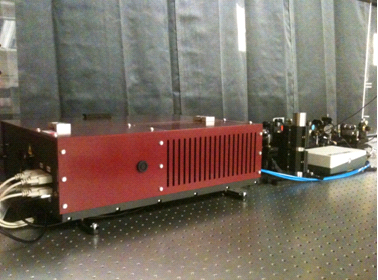 |
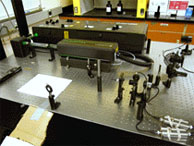
Trestles-Sprout Particle trapping, manipulation and detection are performed on a dedicated setup based on a tunable fs/cw laser system, consisting of a Del Mar Trestles-100M-CW and a Lighthouse Photonics Sprout 10 pump laser. (10W pump power at 532nm). The Trestles allows for both femtosecond pulses and continuous wave pulses to be tuned from 750-850nm with output of about 2W.
|
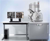 |
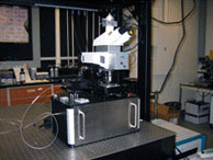
Dual Beam Focused Ion/Electron Beam Nanofabrication Tool For characterization of nanoscale devices and structures, we use a custom-modified Digital Instruments Nanoscope scanning probe microscope. The microscope is used for various types of scanning probe measurements such as atomic force microscopy, scanning capacitance microscopy, and near-field scanning optical microscopy (NSOM). |
|
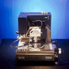
Scanning Force Microscope For characterization of nanoscale devices and structures, we use a custom-modified Digital Instruments Nanoscope scanning probe microscope. The microscope is used for various types of scanning probe measurements such as atomic force microscopy, scanning capacitance microscopy, and near-field scanning optical microscopy (NSOM). |
|
Time-Correlated Single Photon Counting A time-correlated single photon counting setup (TCSPC) is used to carry out studies on single molecules on a chip. Hardware from Picoquant Inc. allows us to cover a wide range of time scales from nanoseconds to seconds.
|
|
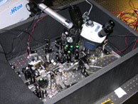
Widely Tunable Femtosecond Laser System Studies of dynamical processes on the picosecond and sub-picosecond scale are done with a Coherent ultrafastlaser system consisting of: MIRA 900-F (femtosecond pulses), MIRA 900-D (femto- and pciosecond pulses), MIRA OPO (Optical parametric oscillator with intracavity frequency doubling), and 9300 SHG frequency doubler. This system delivers ultrashort laser pulses with nearly continuous tunability between 350 and 1300nm, and from 2200-3000nm. This laser system is also integrated with the NSOM to enable optical measurements with simultaneously high spatial and temporal resolution. Support by a Major Research Instrumentation Grant (NSF) and a DURIP Award (ONR) is gratefully acknowledged. |
|
Near-Field Scanning Microscope We use a custom-modified Witec Alpha 300S near-field mciroscope for high reslution optical characterization of various structures. One main focus is the development of a magneto-optical near-field microscope for studies of single nanomagnets in dense arrays. Support by a Major Research Instrumentation (MRI) award from the National Science Foundation is gratefully acknowledged.
|
|
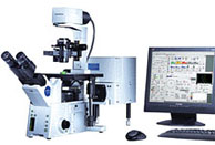
Confocal Microscopy System The Olympus FV1000 is a confocal microscopy system for high resolution scanning imaging down to a few 100nm. Due to its extreme versatility with multiple excitation sources and detectors, we can use it for a variety of applications including picosecond acoustic spectroscopy, measurements of thermoelectric properties of superlattice structures, high-resolution magneto-optic spectroscopy, and time-resolved fluorescence spectroscopy (e.g. FRET) of biomolecules. Support by a DURIP Award (ONR) is gratefully acknowledged. |
|
Magneto-Optical Kerr Spectroscopy This automated setup was built to routinely carry out polarization measurements on magnetic samples using the magneto-optical Kerr effect. Kerr measurements can be taken with angular resolution of 8 mrad and a spatial resolution of 1 mm.
|
|
Waveguide Mode Profiling In order to image the spatial dependence of the optical modes in integrated optical waveguide structures, a high magnification lens in conjunction with CCD detection and dedicated computer control is utilized for measuring the near-field of the optical mode. Diffraction-limited imaging with a resolution in the sub-micron range is possible.
|
|
White Light Source The combination of a femtosecond laser, a hollow-core optical fiber, and a spectrometer allows us to generate femotsecond optical pulses covering most of the visible spectrum for use in waveguide characterization and other measurements.
|

 |